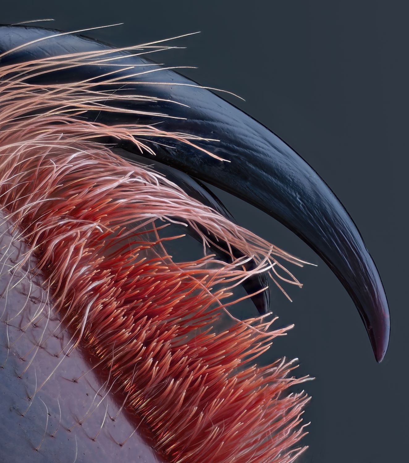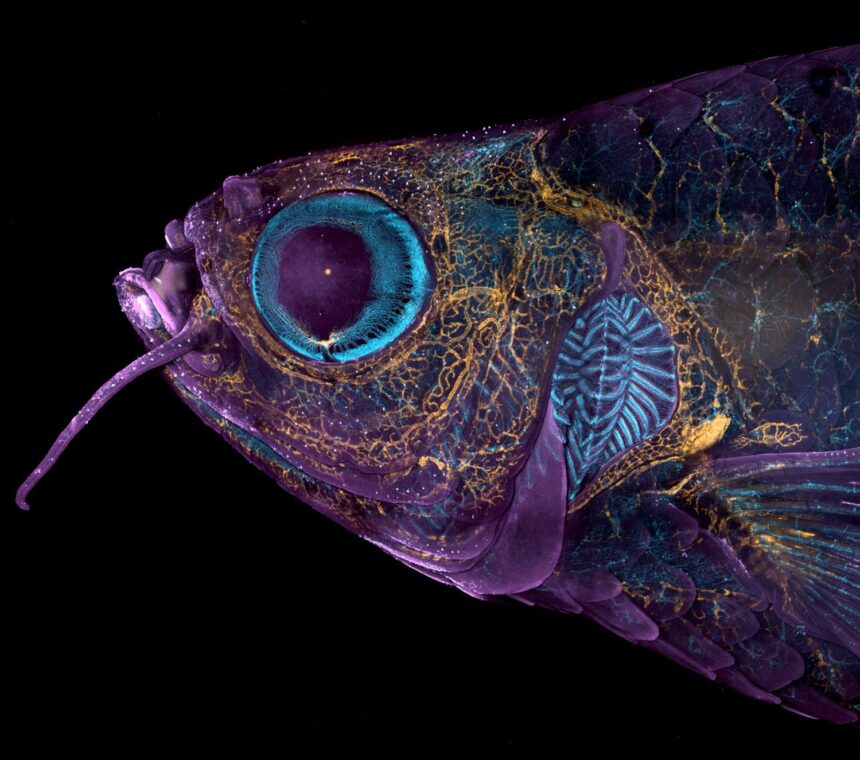
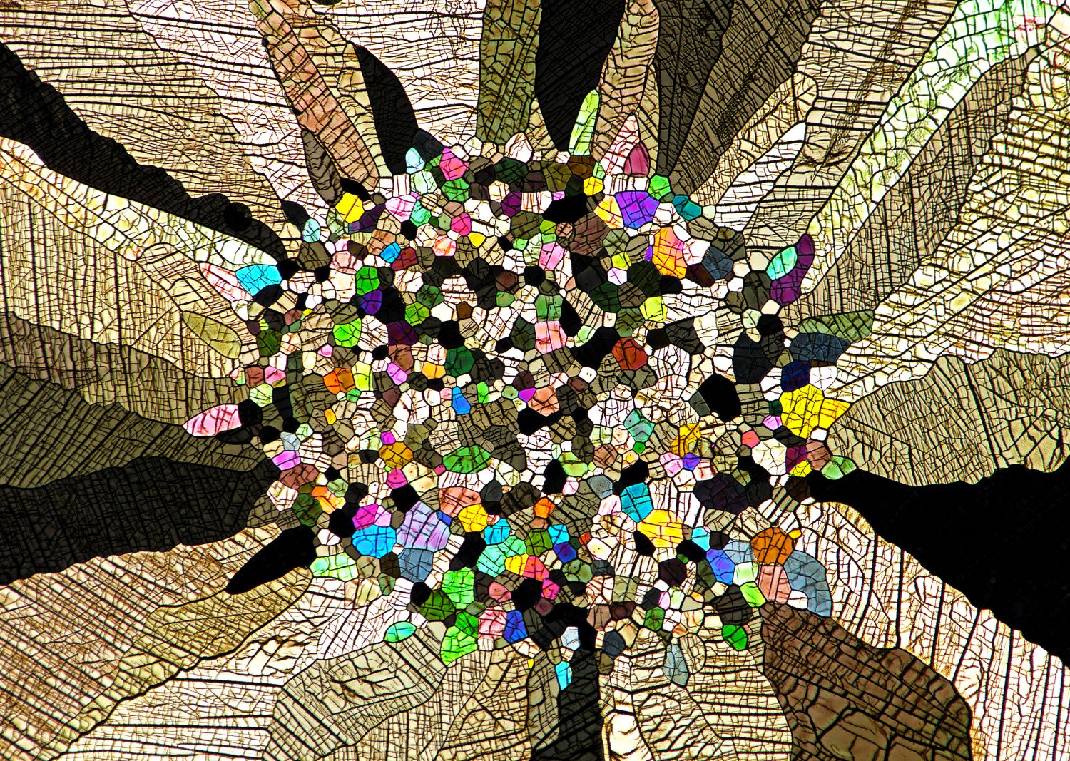
For practically half a century, Nikon’s Small World Photomicrography Competition has celebrated the wonder captured by excessive magnification. This 12 months, the photomicrography contest was stacked: a panel of journalists and scientists chosen winners from 1,900 entries submitted by researchers and photographers in 72 nations. Topics as various as mutant fish, chemical reactions, and a speck of house rock turned artistic endeavors when seen actually, actually up shut.
Above, in first place, is a rodent’s optic nerve head. Blood vessels, every solely 110 microns in diameter, radiate outward just like the fizzing arms of a firework. The yellow star-like shapes surrounding the vessels are astrocytes, mobile helpers that preserve neuronal methods. Imaginative and prescient researchers on the Lions Eye Institute in Perth, Australia—Hassanain Qambari, assisted by Jayden Dickson—imaged the optic disc at 20x magnification as a part of a research of diabetic retinopathy; this situation may cause blindness in folks with diabetes.
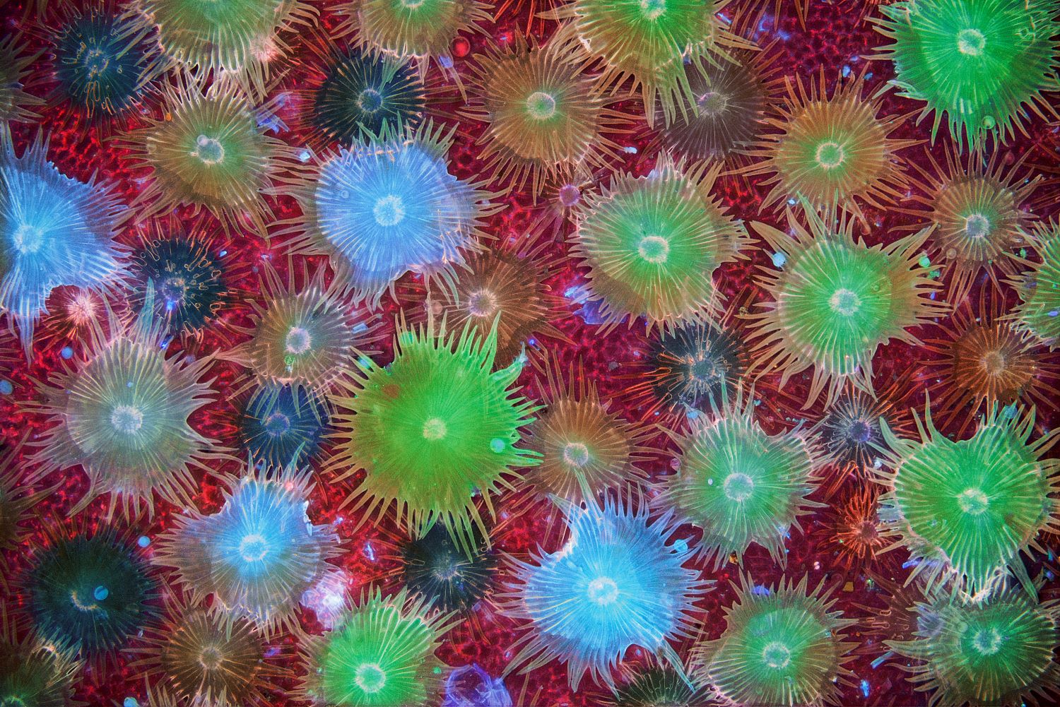
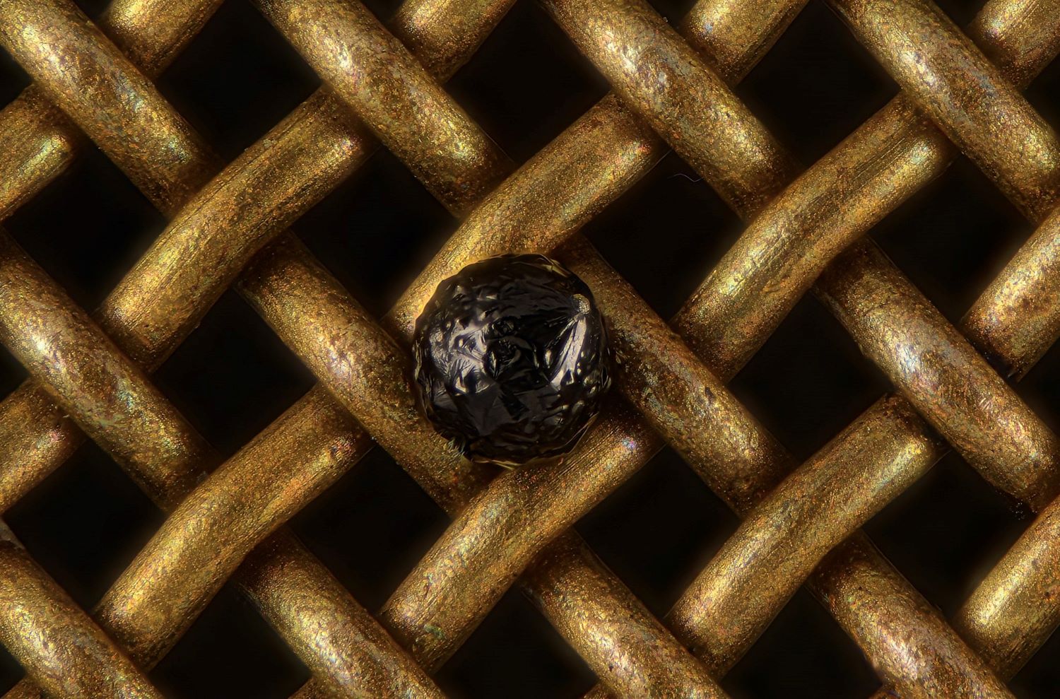
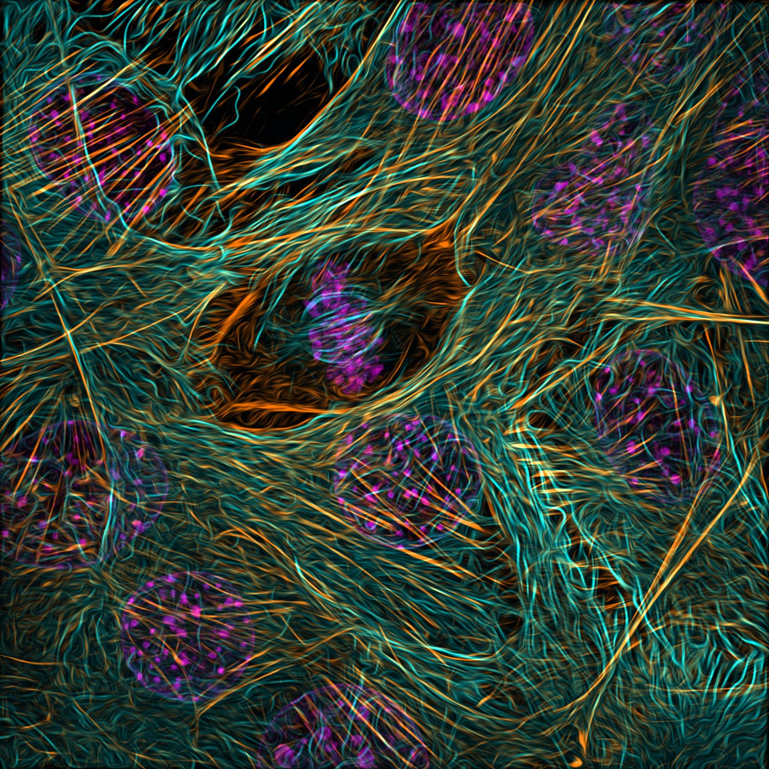
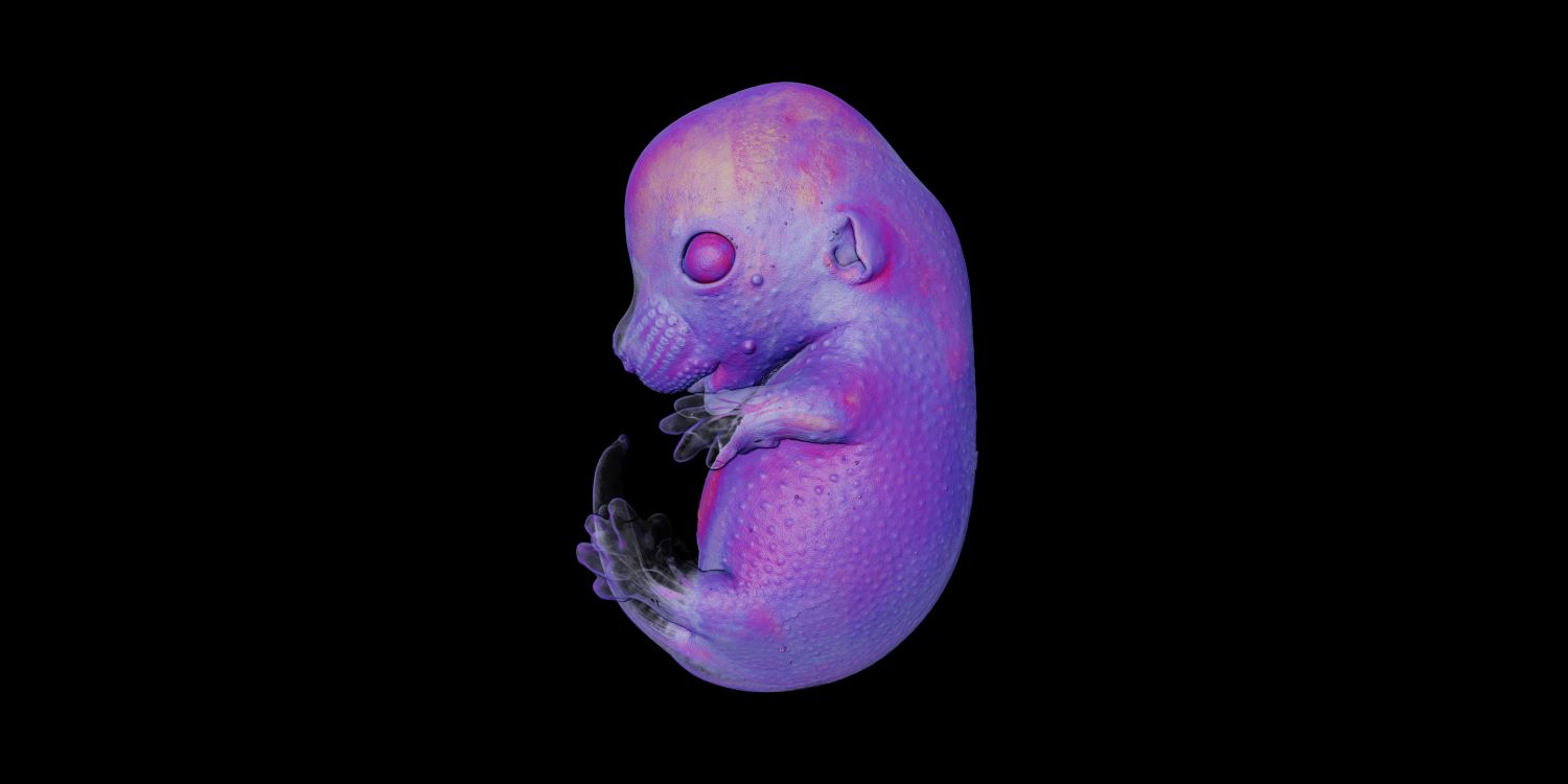
“The visible system is a fancy and extremely specialised organ, with even comparatively minor perturbations to the retinal circulation capable of trigger devastating imaginative and prescient loss,” Qambari mentioned in a information launch. “I entered the competitors as a strategy to showcase the complexity of retinal microcirculation.” Beneath are different prime images, and you may see much more at Nikon’s Small World site.


[Related: 15 remarkable JWST images that reveal the wonders of our vast universe]